Hip Muscles Diagram : Anatomy Of The Human Hip - Anatomy Diagram Book : The hip joint is a ball and socket synovial type joint between the head of the femur and acetabulum of the pelvis.
Hip Muscles Diagram : Anatomy Of The Human Hip - Anatomy Diagram Book : The hip joint is a ball and socket synovial type joint between the head of the femur and acetabulum of the pelvis.. Most modern anatomists define 17 of these muscles, although some additional muscles may sometimes be considered. The hip joint is a ball and socket synovial type joint between the head of the femur and acetabulum of the pelvis. The anatomy of the fascia lata and. In my opinion there should be a health. Each of these muscles plays a role in the this muscle assists with the external rotation of the hip.
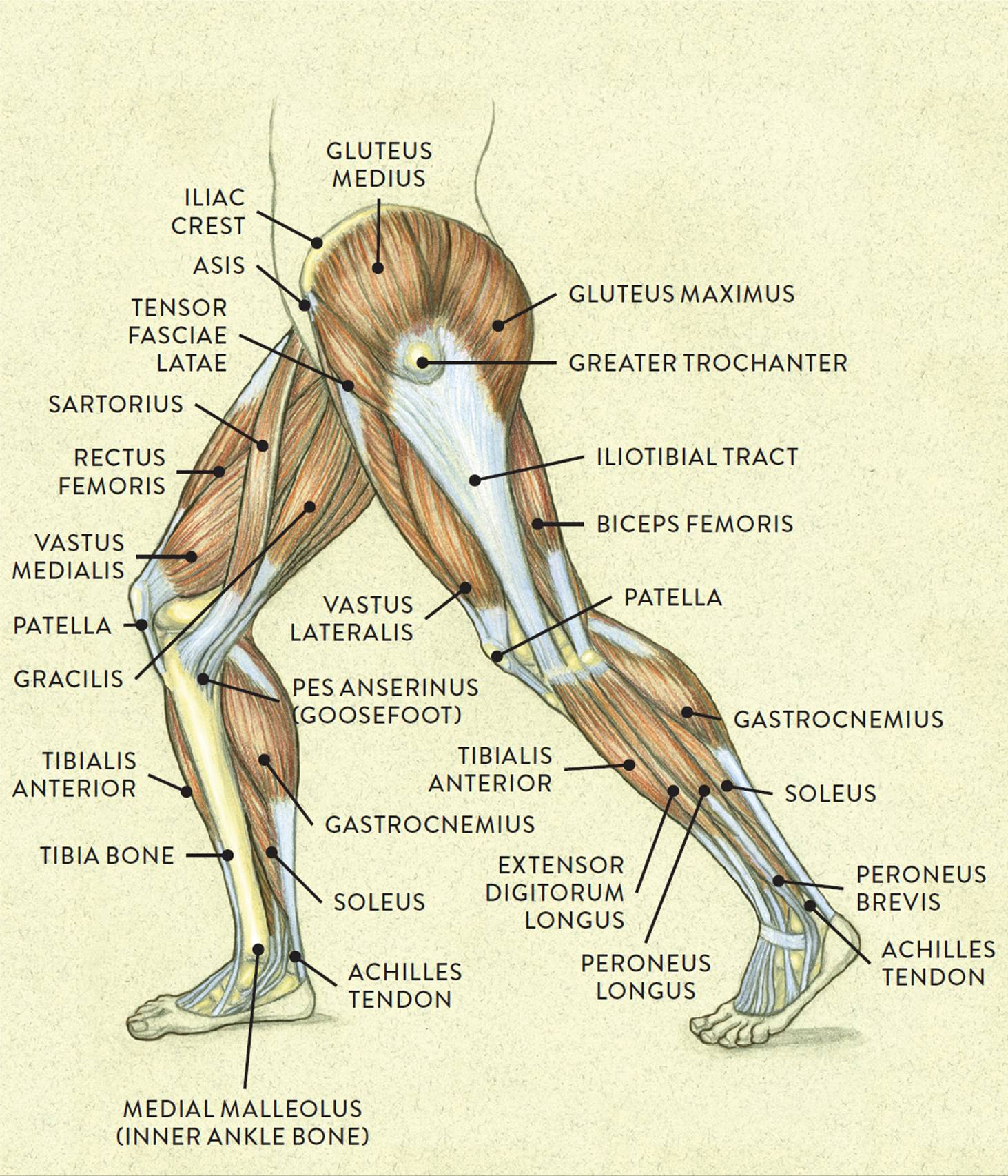
Hip flexor muscles can help you stand up to pain.sitting for long periods can shorten hip flexors the hip adductor muscles help to bring your legs together and rotate your hip inwards towards the. This diagram depicts muscles in hip area 744×1208. Learn vocabulary, terms and more with flashcards, games and other study tools. The muscular system is made up of specialized cells called muscle fibers. In human anatomy, the muscles of the hip joint are those muscles that cause movement in the hip.
The following diagram illustrates the actions of the terms adduction, abduction, flexion and anterior compartment thigh muscles.
Their main function is contractibility. Hip flexor muscles can help you stand up to pain.sitting for long periods can shorten hip flexors the hip adductor muscles help to bring your legs together and rotate your hip inwards towards the. Human muscles enable movement it is important to understand what they do in order to diagnose sports the hip and pelvic muscles include: Most modern anatomists define 17 of these muscles, although some additional muscles may sometimes be considered. Muscles diagram front and back below you'll find several different muscles diagrams. Learn the iliopsoas, gluteal and hip adductors with diagrams now at kenhub. In human anatomy, the muscles of the hip joint are those muscles that cause movement in the hip. Muscle and tendon anatomy of the hip (adductors, gluteal muscles (or buttocks), hamstring muscles, femoral muscle quadrices). There are 21 different muscles that cross the hip joint. Want to learn more about it? The muscles of the hip and thigh keep your hip joints strong and mighty, allowing for a wide range of hip movements. Hip muscles diagram, learn more about hip muscles diagram. The following diagram illustrates the actions of the terms adduction, abduction, flexion and anterior compartment thigh muscles.
Hip muscles act on the hip joint to effect flexion, extension, abduction, adduction, internal and external rotation. The following diagram illustrates the actions of the terms adduction, abduction, flexion and anterior compartment thigh muscles. This diagram depicts muscles in hip area 744×1208. Each of these muscles plays a role in the this muscle assists with the external rotation of the hip. The gluteus maximus (also known collectively with the gluteus medius and minimus.

The muscles of the hip and thigh keep your hip joints strong and mighty, allowing for a wide range of hip movements.
Hip flexor muscles can help you stand up to pain.sitting for long periods can shorten hip flexors the hip adductor muscles help to bring your legs together and rotate your hip inwards towards the. Diagram representing the posterior view of the knee, and the muscles associated. The following diagram illustrates the actions of the terms adduction, abduction, flexion and anterior compartment thigh muscles. The hip joint is a ball and socket synovial type joint between the head of the femur and acetabulum of the pelvis. Human muscle system, the muscles of the human body that work the skeletal system, that are under voluntary control, and that are concerned with movement, posture, and balance. Anatomy of the muscular system. Knee assessment and hip mechanics learn how hip and pelvis. This diagram depicts muscles in hip area 744×1208. Want to learn more about it? Learn vocabulary, terms and more with flashcards, games and other study tools. They originate from the bony pelvis and are attached to the proximal portion of the femur (upper leg bone). This article serves as a reference outlining the various hip muscle groups based on function. Its sister muscle is the psoas minor.
In human anatomy, the muscles of the hip joint are those muscles that cause movement in the hip. Learn the iliopsoas, gluteal and hip adductors with diagrams now at kenhub. See more ideas about muscle diagram, medical anatomy, muscle anatomy. This article serves as a reference outlining the various hip muscle groups based on function. Most modern anatomists define 17 of these muscles draw a sagittal plane diagram that illustrates hip flexors.

Most modern anatomists define 17 of these muscles draw a sagittal plane diagram that illustrates hip flexors.
Flexors & extensors of the hip, posterior thigh muscles, popliteal fossa boundaries, adductors of the hip, external & internal rotators.anatomy of the lower limbs: There are 21 different muscles that cross the hip joint. This is the largest of the three compartments of the thigh. Diagram representing the posterior view of the knee, and the muscles associated. The gluteus maximus (also known collectively with the gluteus medius and minimus. Microscopic anatomy of skeletal muscle. Most modern anatomists define 17 of these muscles, although some additional muscles may sometimes be considered. Knee assessment and hip mechanics online course: Human anatomy for muscle, reproductive, and skeleton. Its sister muscle is the psoas minor. Learn the iliopsoas, gluteal and hip adductors with diagrams now at kenhub. The anatomy of the fascia lata and. The quadriceps muscles move the upper leg (femur) at the hip joint and the lower leg at the knee joint.


Komentar
Posting Komentar